Aim: To calculate size, shape and position of mental foramen. Materials & Methods: 50 dry human mandibles of either gender (20- females, 30- males) were included. The position of mental foramen in horizontally was calculated based on classification proposed by Bokhari. The vertical position was divided into six types using the modified Ngeow and Yuzawati criteria. Size was measured both vertically and horizontally with the help of vernier caliper and expressed as mean. Results: Most common horizontal position was II seen in both males (left- 56%, right- 50%) and females (left- 60%, right- 58%). Most common vertical position was 2 seen in males (left- 62%, right- 65%) and females (left- 55%, right- 52%). Most common shape was oval seen in both genders (males- 68% left, 62% right) and (females- 70% left, 72% right). A significant difference was observed \((P<0.05)\). Conclusion: Variation in shape, size and position was observed both males and females, however, most common shape found to be oval and position was II horizontally and 2 vertically in both genders.
Mental foramen is an opening present on lateral aspect of mandible. These are to in number on right side and one on left side [1]. Here, the mandibular nerve unites with mental nerve and may continue as incisive nerve. It also caries mental vessels [2,3]. The location of mental foramen vary person to person and with different age group. The position of mental foramen both vertically and horizontally has been classified by various authors [4,5]. In children before eruption of teeth, it is normally present near alveolar crest, Similarly, in geriatric population due to continuous bone resorption it is close to the crestal bone [6]. Normally, it is present below premolars, the position may be between first and second premolar, anterior to first premolar, anterior to second premolar or anterior to first molar [7]. Vertically it is classified into 6 positions based on its occurrence within 2 mm of root of first premolar and second premolar. The occurrence of accessory mental foramen is not uncommon, if present, it usually lies below first molar [8].
Mental nerve innervates the lower lip, labial mucoperiosteum of the ipsilateral lower incisors, canine and premolars. The size and shape also vary. It is either oval, irregular or circular shape. Size may vary from 2-4 mm [9]. The thorough knowledge of position, size and shape of mental foramen is of great value as various surgical procedures such as insertion of dental implant, orthognathic surgeries, dental filling are frequently done in mandible [10]. Sometimes, surgical procedure performed on mandible can lead to paraesthesia of lower lip and chin if the position of mental foramen is not taken into consideration. Therefore, in order to prevent such iatrogenic injuries, the knowledge its exact location is of paramount importance [11]. Considering this, we attempted this study on 50 dry human mandibles to calculate size, shape and position of mental foramen.
The position of mental foramen in horizontally was calculated based on classification proposed by Bokhari. Position I: mesial to the first premolar; Position II: between the first and second premolars; Position III: distal to the second premolars. The radiographic vertical position was divided into six types using the modified Ngeow and Yuzawati criteria. Position 1: when it is present more than 2 mm inferior to the apex of the first premolar. Position 2: when it is present more than 2 mm inferior to the apex of the second premolar. o Position 3: when it is less than 2 mm inferior or at the apex of the first premolar. o Position 4: when it is 2 mm inferior or at the apex of the second premolar. o Position 5: when it is positioned superior to the apex of the first premolar. o Position 6: when it is present superior to the apex of the second premolar. All these finding s were measured following radiographic analysis done on OPG radiograph taken with machine Allengers following al standardized parameters. Size was measured both vertically and horizontally with the help of vernier caliper and expressed as mean. Results of the present study after recording all relevant data were subjected for statistical inferences using chi- square test. The level of significance was significant if p value is below 0.05 and highly significant if it is less than 0.01.
| Dimension (mean) (mm) | Male | Female | P value | ||
|---|---|---|---|---|---|
| Left | Right | Left | Right | ||
| Vertical | 2.90 | 2.88 | 2.82 | 2.86 | Non- significant, >0.05 |
| Horizontal | 3.15 | 3.12 | 3.20 | 3.21 | Non- significant, >0.05 |
It was observed that mean vertical dimension of mental foramen in males left side was 2.90 mm and on right side was 2.88 mm and in females left side was 2.82 mm and on right side was 2.86 mm. Horizontal dimension was 3.15 mm in males left side and 3.12 mm on right side and 3.20 mm in females left side and 3.21 mm in right side. A non- significant difference was observed (P> 0.05) (Table1 ).
| Position | Category | Male | Female | P value | ||
|---|---|---|---|---|---|---|
| Left | Right | Left | Right | |||
| Horizontal | I | 30% | 39% | 22% | 25% | >0.05 |
| II | 56% | 50% | 60% | 58% | <0.05 | |
| III | 14% | 11% | 18% | 20% | >0.05 | |
| Vertical | 1 | 5% | 3% | 2% | 5% | <0.05 |
| 2 | 62% | 65% | 55% | 52% | >0.05 | |
| 3 | 3% | 4% | 14% | 11% | >0.05 | |
| 4 | 30% | 28% | 22% | 24% | <0.05 | |
| 5 | 0 | 0 | 7% | 8% | >0.05 | |
| 6 | 0 | 0 | 0 | 0 | – | |
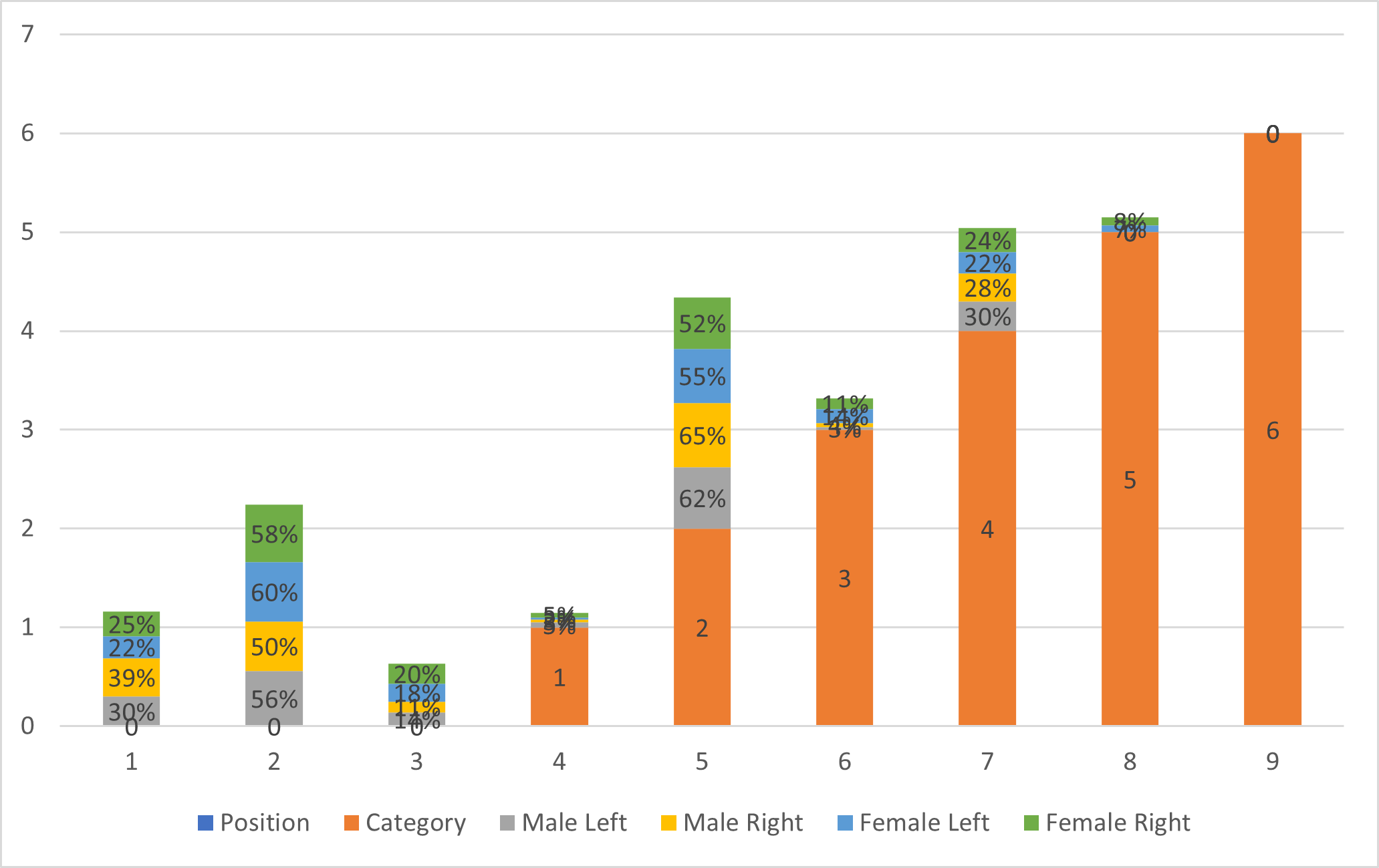
Most common horizontal position was II seen in both males (left- 56%, right- 50%) and females (left- 60%, right- 58%). Most common vertical position was 2 seen in males (left- 62%, right- 65%) and females (left- 55%, right- 52%). A significant difference was observed (P< 0.05) (Table 1, Figure 1).

I am raw html block.
Click edit button to change this
| Shape | Male | Female | P value | ||
|---|---|---|---|---|---|
| Left | Right | Left | Right | ||
| Circular | 12% | 20% | 14% | 13% | <0.05 |
| Oval | 68% | 62% | 70% | 72% | >0.05 |
| Irregular | 20% | 18% | 16% | 15% | <0.05 |
Most common shape was oval seen in both genders (males- 68% left, 62% right) and (females- 70% left, 72% right). A significant difference was observed (P< 0.05) (Table 3).
Our study found higher vertical dimension in males compared to females. In males on left side was 2.90 mm and on right side was 2.88 mm and in females left side was 2.82 mm and on right side was 2.86 mm. Horizontal dimension was 3.15 mm in males left side and 3.12 mm on right side and 3.20 mm in females left side and 3.21 mm in right side. Bello [17] in their study took 320 orthopantomograms of subjects and observed that most of the foramina analysed were horizontally positioned between the mandibular first and second premolars (65.9%) and vertically positioned greater than 2 mm below the apex of the second mandibular premolars. The average vertical dimension and horizontal dimension of the foramen is 2.87 mm and 3.56 mm respectively with 55.2% of the foramen analysed being ovoid in shape. Asymmetrical mental foramina were seen in 164 subjects (51.3%) while 156 subjects had symmetrical mental foramina (48.7%). A study done on a Turkey population reported the HD to be 2.93 mm on the right side and to be 3.14 mm on the left side; the vertical Diameter (VD) was 2.38 mm on the right side and it was 2.64 mm on the left side.
Our study showed that most common horizontal position was II seen in both males ie. left- 56%, right- 50% and females ie. left- 60%, right- 58%. Most common vertical position was 2 seen in males ie. left- 62%, right- 65% and females ie. left- 55%, right- 52%. Cabanillas [18] in their study using 180 cone beam CTs analyzed the distance between the upper and lower cortical areas of the mental foramen to the alveolar crest and the mandibular basal bone respectively, as well as the location, shape, size and presence of accessory holes. The mean of the upper cortical area in relation to the alveolar crest was 15.00 mm and the mean of the lower cortical area to the mandibular basal bone was 13.75 mm. The most frequent location was the longitudinal axis of the second premolar (44.4% right side and 47.2% left side). The predominant shape was oval and the size was in the range of 2.00 mm to 2.99 mm. Accessory holes were present in 55.5% of cases.
It was shown in our results that most common shape was oval seen in 68% left and 62% right side in males and 70% left and 72% right in females. Udhaya et al., [19]. conducted a study on 90 adult dry human msandibles to locate position and found that the mental foramen was located at the level of the root of the 2nd premolar, midway between the inferior margin and the alveolar margin of the mandible. Most of the mental foramina were oval in shape. The orientation of the foramen was postero-superior in 83% of the mandibles. The accessory foramens were noted in five mandibles. Our study did not report any occurrence of accessory foramen in either side of genders. Cag Irankaya et al., [20]. reported AMFs below the 1st molar.