
Tuberculosis (TB), caused by Mycobacterium tuberculosis (Mtb), continues to be one of the leading infectious causes of mortality worldwide. According to the World Health Organization, tuberculosis continues to affect millions of people annually, while the emergence and spread of drug-resistant strains further complicate disease control and treatment [1]. The effectiveness of first-line drugs such as isoniazid and rifampicin is compromised when resistant strains emerge, including multidrug-resistant TB (MDR-TB) and extensively drug-resistant TB (XDR-TB) [2]. This persistent threat has necessitated the development of new chemotherapeutic agents with improved activity and better resistance profiles.
Schiff bases, first described by Hugo Schiff in 1864, have attracted sustained interest in medicinal chemistry because their imine functionality permits a broad range of structural modifications and biological investigations [3]. Several published reviews describe antibacterial, antifungal, antiviral, and other biological activities among individual Schiff-base derivatives and their metal complexes; such activities must be established separately for each newly synthesized compound [4]. The heterocyclic substitution pattern and the availability of nitrogen- and oxygen-containing groups can influence coordination chemistry and molecular interactions, supporting further investigation of these structures without implying established drug efficacy [5]. Indole-containing compounds are often investigated in medicinal chemistry because their ring system permits extensive structural diversification. The review by da Silva et al. describes the broad antimicrobial potential of Schiff bases, although it does not independently verify any particular indole compound as a tuberculosis inhibitor [6]. Related indole-containing hydrazides and hydrazones have shown experimentally measured activity against selected tuberculosis strains, providing a chemical rationale for exploring additional indole-containing scaffolds [7]. For instance, Verma et al. [8] reviewed the biological properties of hydrazones, including their reported antitubercular activity. Because that publication is a review of hydrazones rather than an experimental report on indole-derived Schiff bases, it should not be presented as direct evidence of inhibition by the compounds examined here. For example, Varma et al. synthesized Schiff bases and 1,3,4-oxadiazole derivatives and assessed their activity against M. tuberculosis H37Rv, demonstrating that these chemical families have been investigated experimentally, although their compounds differ from the present derivatives [9].
Amino-acid-derived Schiff bases have attracted attention because their polar functional groups and variable side chains provide additional options for altering molecular properties [10]. Depending on the particular side chain, amino-acid-derived groups may influence solubility, introduce additional hydrogen-bonding sites, and alter lipophilicity, although improvement in pharmacokinetic properties cannot be assumed without testing [10]. A review by Kumari et al. summarizes the synthesis and biological investigations of amino-acid-derived Schiff bases and their metal complexes; it does not establish binding to the particular tuberculosis protein target considered in the present study [10]. Recent advances in computational chemistry have facilitated the use of molecular docking to predict ligand–protein interactions and identify promising lead candidates before in vitro experimentation. Structure-based screening has been applied to validated tuberculosis targets such as InhA (the enoyl-acyl carrier protein reductase) and DprE1, but computational docking requires careful target selection and subsequent biological validation [11].
Although previous studies have established the broad biological potential of Schiff bases, important limitations remain in their development as antitubercular candidates. Heterocyclic Schiff bases offer structural diversity and multiple hydrogen-bonding and hydrophobic interaction sites, but reported biological activities are highly dependent on the nature and position of the substituents, and direct comparison among different scaffolds is often difficult because different studies employ different mycobacterial strains, protein targets, experimental conditions, and computational protocols. Indole-containing Schiff bases have demonstrated promising antimycobacterial properties, while amino-acid-derived Schiff bases may provide additional advantages through their hydrogen-bonding capacity, polarity, and structural flexibility. However, many reported investigations remain at the preliminary stage, with computational docking or basic antimicrobial screening not always being followed by comprehensive in vitro, cytotoxicity, pharmacokinetic, or in vivo validation. Similarly, favorable docking scores should be interpreted cautiously because they represent predicted ligand–protein interactions rather than direct evidence of biological inhibition. These limitations indicate a need for systematically designed Schiff-base derivatives in which structural modifications can be correlated with predicted binding interactions and subsequently validated experimentally. In this context, the combination of a 2-phenylindole pharmacophore with amino-acid-derived functionality represents a rational strategy for generating structurally diverse candidates. Nevertheless, comparative evaluation of glutamine-, histidine-, and valine-derived derivatives against the same M. tuberculosis target remains insufficiently explored. The present study was therefore designed to address this gap by synthesizing three related derivatives and comparing their reported docking interactions with a structure identified as 6PUV against those obtained for isoniazid. The identity of the deposited 6PUV structure must nevertheless be reconciled with the stated tuberculosis-specific objective.
The literature cited in this study was used to establish the background and the scientific rationale for the design of the present heterocyclic Schiff bases. Previous reports on Schiff bases, indole-containing derivatives, amino-acid-derived Schiff bases, and computational investigations of potential Mycobacterium tuberculosis inhibitors were considered in formulating the research hypothesis and selecting the molecular scaffold and target. The present work, however, is an experimental and computational investigation rather than a systematic or scoping literature review, and its background references were not collected through a formal evidence-synthesis protocol. Accordingly, no formal systematic literature-search protocol, predefined study-selection criteria, quantitative study screening, or PRISMA-based selection process was undertaken. The cited literature is intended to provide the scientific context for the present synthesis, characterization, and molecular docking study.
In this study, three heterocyclic Schiff bases were synthesized by condensing 2- phenylindole-3- carboxaldehyde with the amino acids L-glutamine, L-histidine, and valine. The design rationale combines the bioactive indole core with amino-acid functionality, aiming to enhance binding affinity and improve drug-like properties. Structural characterization was performed using spectroscopic methods to investigate product formation and assess the identities of the derivatives; quantitative purity was not established solely through these techniques. To explore predicted ligand–protein interactions, molecular docking studies were described for PDB entry 6PUV. The archive identifies this accession as a human macrophage galactose C-type lectin domain, not an M. tuberculosis protein; therefore, the target designation and its relevance to tuberculosis require correction or experimental clarification. Predicted binding scores were also compared with those generated for the standard antituberculosis drug isoniazid, although its use as a receptor-specific positive control was not established. The reported docking results yielded more negative scores for all three Schiff bases than for isoniazid and suggested possible hydrogen-bonding and hydrophobic contacts; these calculations cannot establish increased complex stability or biological inhibition. Given these findings and the growing body of evidence supporting Schiff bases as promising antitubercular agents, this study provides a preliminary basis for examining structure-dependent interactions and for planning future in vitro and in vivo evaluations after the receptor assignment has been resolved.
Despite the considerable interest in Schiff bases and indole-based heterocyclic compounds, several limitations remain in their development as potential antitubercular agents. Previous studies have primarily investigated diverse Schiff-base frameworks, individual heterocyclic substituents, or established Mycobacterium tuberculosis targets such as InhA and DprE1, while comparatively limited attention has been given to integrating a 2-phenylindole pharmacophore with amino-acid-derived Schiff-base functionality and evaluating structurally related derivatives against a common protein target. In addition, many computational investigations provide useful predictions of ligand–protein interactions but require subsequent experimental validation to establish actual antimycobacterial efficacy, selectivity, and pharmacological suitability. The present study addresses this research gap by designing and synthesizing three Schiff bases derived from 2-phenylindole-3-carboxaldehyde and the amino acids L-glutamine, L-histidine, and valine, followed by spectroscopic characterization and comparative molecular docking against the structure designated by PDB ID 6PUV, subject to verification of the receptor identity. The significance of the work lies in establishing a structure-dependent comparison of these amino-acid-derived derivatives and identifying molecular features associated with their predicted binding behavior. In particular, the study provides a preliminary basis for selecting promising candidates for subsequent in vitro antimycobacterial testing, cytotoxicity assessment, pharmacokinetic investigation, and structure–activity relationship optimization.
The heterocyclic Schiff base derivatives were prepared through a condensation reaction between 2-phenylindole-3-carboxaldehyde and the amino acids L-glutamine, L-histidine, and valine in an ethanolic medium under reflux. Initially, 2-phenylindole-3-carboxaldehyde (1 mmol) was dissolved in 10–15 mL of ethanol in a round-bottom flask with continuous magnetic stirring. In parallel, equimolar amounts of each amino acid were dissolved either in ethanol or in a minimal amount of water for highly polar amino acids. Each amino acid solution was then added dropwise to the aldehyde solution under constant stirring.
To promote condensation and facilitate imine (Schiff base) formation, a few drops of glacial acetic acid were introduced as a catalyst, maintaining the reaction mixture at a pH of approximately 5–6, using the acid-catalyzed condensation approach described in the general Schiff-base synthetic literature [6]; the specific conditions and quantities described here are those reported for the present experiments. The reaction mixture was subsequently heated under reflux at a reported temperature of 60–70 \(^\circ\)C for 4–6 hours. Reaction progress was monitored by thin-layer chromatography (TLC) using an ethanol:chloroform (3:7) mobile phase as specified in the experimental description.
Upon completion, the mixture was cooled to room temperature and then placed in an ice bath to promote product precipitation. If crystallization did not occur, the solvent was removed under reduced pressure using a rotary evaporator. The resulting solid was filtered under vacuum, washed thoroughly with cold ethanol or diethyl ether to remove unreacted residues, and dried in a vacuum oven at 50 \(^\circ\)C to obtain the reported Schiff base products. The synthesized derivatives were characterized by Fourier-transform infrared (FTIR) spectroscopy and proton/carbon-13 nuclear magnetic resonance (\(^1\)H/\(^{13}\)C NMR) spectroscopy to support the proposed molecular structures; chromatographic and other purity evidence should be considered separately. The resulting compounds were described as follows: (E)-2-((2-phenyl-1H-indol-3-yl)methyleneamino)-4-carbamoylbutanoic acid (L-glutamine derivative), (E)-2-((2-phenyl-1H-indol-3-yl)methyleneamino)-3-(1H-imidazol-4-yl)propanoic acid (L-histidine derivative), and 2-((2-phenyl-1H-indol-3-yl)methyleneamino)-3-methylbutanoic acid (valine derivative). These Schiff bases were selected to compare the effects of three chemically distinct amino-acid-derived side chains on the structure and predicted interaction patterns of a shared 2-phenylindole scaffold. Previous reviews describe antimicrobial properties of other Schiff-base derivatives, but the biological activities of the present three compounds have not been demonstrated [4].
Molecular docking studies were described to estimate the predicted binding scores and molecular interactions of the synthesized Schiff bases with the receptor file assigned PDB ID 6PUV. The official Protein Data Bank identifies 6PUV as the human macrophage galactose C-type lectin domain rather than a Mycobacterium tuberculosis H37Rv protein [12]; consequently, the correct receptor file must be confirmed before the docking results can be linked to an antimycobacterial target. The ligand set included the three synthesized compounds and the antitubercular drug isoniazid as a computational comparator. Ligand structures were either drawn using ChemDraw Ultra 12.0 (PerkinElmer Inc., USA) or retrieved from the PubChem database [13]. The 3D structure identified by the authors as protein 6PUV was downloaded from the Protein Data Bank [12, 14]. Docking simulations were carried out using AutoDock Vina [15], while visualization of docking results was performed using PyMOL (Schrödinger LLC) and BIOVIA Discovery Studio Visualizer. Ligand optimization and energy minimization were conducted using Avogadro [16], and file format conversions were performed with Open Babel [17].
Ligand structures were imported into Avogadro for geometry optimization using the MMFF94 force field [18]. The optimized structures were converted to PDB format via Open Babel. Ligand preparation for docking was completed using AutoDockTools (ADT) 1.5.7, during which polar hydrogens were added, Gasteiger charges were assigned, and rotatable bonds were defined. The final ligand structures were saved in PDBQT format for docking [19].
The deposited structure identified as 6PUV was processed using PyMOL by removing water molecules and any non-essential heteroatoms or bound ligands. The cleaned structure was then imported into AutoDockTools, where polar hydrogens were added and Kollman charges were assigned. The final receptor file was saved in PDBQT format for docking simulations [19]. Figure 1 shows the receptor structure used for the reported docking analysis. The coordinate file was obtained from the Protein Data Bank and prepared as described in the materials and methods section; the organism and protein identity associated with the accession must be checked against the original docking inputs.
The putative binding region was described as being selected using earlier literature and information associated with the deposited structure. PyMOL was used to examine the receptor geometry [20]; this software reference does not independently validate the selected binding site. Grid box dimensions were set in AutoDockTools to fully encompass the binding region. Grid-center coordinates (x, y, z) were aligned with the selected binding region, and the grid dimensions were adjusted to permit conformational exploration. The numerical grid-center coordinates and box dimensions were not reported and should be supplied to support reproducibility. A configuration file (config.txt) containing docking parameters—exhaustiveness = 8, number of modes = 9, and energy range = 3 kcal/mol—was generated following Trott & Olson [15].
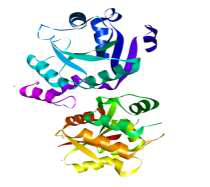
Docking was performed in a Linux environment using the prepared ligand and receptor PDBQT files and the configuration file. Each ligand was docked independently, and the log files were used to extract binding affinity values and docking poses.
Docked complexes were analyzed to determine binding affinity (\(\Delta\)G, kcal/mol) and molecular interactions. The best pose corresponded to the conformation with the lowest binding energy. Visualization using PyMOL and Discovery Studio Visualizer enabled examination of hydrogen bonding, hydrophobic interactions, and \(\pi\)–\(\pi\) stacking interactions [21]. RMSD values were described as being calculated to compare docked poses with a reference conformation; however, neither the numerical RMSD results nor the reference-pose alignment procedure was provided, limiting independent assessment of this validation step.
Docking scores and interaction profiles were considered when comparing the compounds, but these predictions alone cannot establish either complex stability or inhibitory activity. The compounds demonstrating the most favorable binding energies and the highest number of stabilizing interactions were considered promising candidates. The scores were compared with the result obtained for isoniazid and discussed in the context of tuberculosis drug discovery [22]. This comparison is not a docking-protocol validation, especially while the receptor identity remains unresolved. For InhA, native isoniazid is a prodrug and is not equivalent to the activated INH–NAD inhibitor; the choice of a comparator must be justified for the authenticated target [11].
The condensation of 2-phenylindole-3-carboxaldehyde with the amino acids L-glutamine, L-histidine, and valine afforded three novel heterocyclic Schiff base derivatives. The reactions were carried out under reflux in ethanol with glacial acetic acid as a catalyst, which enhanced the electrophilicity of the aldehyde and facilitated the formation of the imine linkage (–C=N–). The resulting Schiff bases were isolated as crystalline solids, and their structures were assessed using FTIR, \(^1\)H/\(^{13}\)C NMR, and mass spectrometry. The reported data were interpreted as supporting imine formation and the proposed molecular structures; however, the assigned molecular formulas and mass-to-charge values should be rechecked for consistency.
The synthesized derivatives were obtained in moderate to high yields, ranging from 72% to 85%, with the Valine-derived Schiff base exhibiting the highest yield (85%), followed by the histidine derivative (78%), and the glutamine derivative yielding 72%. The differences in yield can be attributed to the varying steric and electronic effects of the amino acid side chains, which influence the condensation efficiency with the aldehyde. Overall, the method proved efficient for the synthesis of structurally diverse Schiff bases with potential biological activity. The condensation routes used to prepare the three heterocyclic Schiff-base derivatives from 2-phenylindole-3-carboxaldehyde and L-glutamine, L-histidine, or L-valine are illustrated in Figure 2, with the reaction conditions described in the materials and methods section.
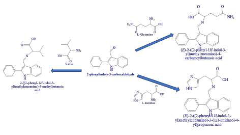
The synthesized Schiff bases were obtained as crystalline solids with colors ranging from pale yellow to light brown, reflecting differences in their conjugated systems and hydrogen bonding interactions. The melting points, determined using a digital melting point apparatus, were distinct for each derivative: the Glutamine Schiff base melted at 195–198 \(^\circ\)C, the histidine derivative at 210–213 \(^\circ\)C, and the valine derivative at 180–183 \(^\circ\)C. All products exhibited higher melting points compared to the starting aldehyde (158–160 \(^\circ\)C), indicating the formation of more stable conjugated systems due to the imine (C=N) linkage. The observed differences in melting points may reflect differences in hydrogen bonding, steric effects, and intramolecular interactions, with the histidine derivative showing the highest melting point, likely due to additional \(\pi\)–\(\pi\) stacking interactions and intramolecular hydrogen bonding involving the imidazole ring. The isolated yields ranged from 72% to 85%, with the valine derivative showing the highest yield (85%), followed by the histidine derivative (78%) and the glutamine derivative (72%), suggesting that steric and electronic effects of the amino acid side chains influenced the condensation efficiency.
Thin-layer chromatography (TLC) was used to monitor reaction progress and to make a qualitative assessment of the resulting products. The aldehyde precursor displayed an Rf value of 0.62, whereas the Schiff base derivatives showed lower Rf values due to their increased polarity: 0.48 for the glutamine derivative, 0.50 for the histidine derivative, and 0.55 for the valine derivative. The trend in Rf values correlates with the polarity of the amino acid side chains; the glutamine derivative, with polar amide (-CONH\(_2\)) and carboxyl (-COOH) groups, exhibited the lowest Rf value, reflecting stronger interactions with the stationary phase, whereas the valine derivative, possessing a nonpolar aliphatic side chain, displayed the highest Rf value. Overall, the TLC results were consistent with changes in chromatographic behavior following condensation under the selected solvent system (ethanol:chloroform, 3:7). However, retention-factor differences alone do not confirm molecular identity or establish quantitative product purity.
The compound (E)-2-((2-phenyl-1H-indol-3-yl)methyleneamino)-4-carbamoylbutanoic acid was obtained as a yellowish crystalline solid. The IR spectrum (KBr) displayed characteristic absorption bands at 3320 cm\(^{-1}\) corresponding to the indole N–H stretch, along with another broad band at 3255 cm\(^{-1}\) attributable to the amide N–H group. Aromatic C–H stretching appeared at 3050 cm\(^{-1}\), while aliphatic C–H vibrations were observed at 2924 cm\(^{-1}\). Strong carbonyl absorptions were noted at 1708 cm\(^{-1}\) for the carboxylic acid and 1651 cm\(^{-1}\) for the amide group. The imine (C=N) stretch appeared prominently at 1610 cm\(^{-1}\), accompanied by aromatic C=C stretching at 1460 cm\(^{-1}\) and characteristic benzene out-of-plane bending at 755 and 700 cm\(^{-1}\).
The \(^1\)H NMR spectrum (400 MHz, DMSO-d\(_6\)) showed a singlet at \(\delta\) 12.01 ppm corresponding to the carboxylic acid proton, followed by a singlet at \(\delta\) 10.98 ppm attributed to the indole NH. The azomethine proton (HC=N) resonated as a sharp singlet at \(\delta\) 8.65 ppm. Multiplets in the region \(\delta\) 7.89–7.22 ppm accounted for nine aromatic protons from the indole and phenyl rings. The amide NH appeared as a singlet at \(\delta\) 6.95 ppm. The aliphatic region exhibited a triplet at \(\delta\) 3.82 ppm (1H) for the methine proton adjacent to the imine carbon, a triplet at \(\delta\) 2.42 ppm (2H) corresponding to the CH\(_2\) group adjacent to the amide carbonyl, and a multiplet between \(\delta\) 2.12–1.98 ppm for the remaining CH\(_2\) group.
The \(^{13}\)C NMR spectrum (100 MHz, DMSO-d\(_6\)) displayed resonances at \(\delta\) 174.8 and 170.2 ppm, assigned to the carboxylic acid and amide carbonyl carbons, respectively. The imine carbon appeared at \(\delta\) 161.3 ppm. Aromatic carbons of the indole and phenyl rings were observed between \(\delta\) 137.4 and 110.5 ppm. Signals at \(\delta\) 56.1, 38.4, and 30.8 ppm corresponded to the methine carbon adjacent to the imine and the two aliphatic CH\(_2\) carbons of the amino-acid-derived side chain. Mass spectrometric analysis (ESI-MS) showed a molecular ion peak at \(m/z\) 350.2 [M+H]\(^+\), consistent with the calculated value of 350.14 for C\(_{20}\)H\(_{19}\)N\(_3\)O\(_3\). Fragmentation peaks at \(m/z\) 332, 304, 258, and 130 indicated cleavage of the imine bond and breakdown of the indole moiety.
The compound (E)-2-((2-phenyl-1H-indol-3-yl)methyleneamino)-3-(1H-imidazol-4-yl)propanoic acid was obtained as a pale-yellow solid with a melting-point range of 210–213 \(^\circ\)C, as reported in the physical characterization results. The IR spectrum (KBr) exhibited a broad absorption band at 3335 cm\(^{-1}\) corresponding to overlapping N–H stretching vibrations from both the indole and imidazole NH groups. A strong O–H stretching signal of the carboxylic acid appeared at 3238 cm\(^{-1}\). Aromatic C–H stretching bands were observed around 3052 cm\(^{-1}\), while aliphatic C–H stretching occurred at 2922 cm\(^{-1}\). The carboxylic acid C=O stretch appeared as a strong band at 1715 cm\(^{-1}\), and the imine (C=N) functional group showed a characteristic peak at 1618 cm\(^{-1}\). Additional signals included aromatic C=C stretching at 1472 cm\(^{-1}\) and out-of-plane C–H bending of the benzene ring at 752 and 702 cm\(^{-1}\).
The \(^1\)H NMR spectrum (400 MHz, DMSO-d\(_6\)) displayed a broad singlet at \(\delta\) 12.03 ppm corresponding to the COOH proton, followed by a singlet at \(\delta\) 11.02 ppm for the indole NH. The imidazole NH appeared downfield at \(\delta\) 8.97 ppm. The azomethine proton (HC=N) resonated as a sharp singlet at \(\delta\) 8.72 ppm. Aromatic protons of the indole and phenyl moieties appeared as multiplets in the region \(\delta\) 7.92–7.18 ppm (9H). The imidazole ring protons were observed at \(\delta\) 7.62 and 7.05 ppm as two distinct singlets. The aliphatic methine proton adjacent to the imine carbon appeared at \(\delta\) 4.12 ppm as a doublet (1H), while the CH\(_2\) protons of the amino-acid-derived chain resonated as multiplets in the region \(\delta\) 3.12–2.84 ppm (2H).
The \(^{13}\)C NMR spectrum (100 MHz, DMSO-d\(_6\)) showed characteristic signals at \(\delta\) 175.6 ppm corresponding to the carboxylic acid carbonyl and at \(\delta\) 161.8 ppm for the azomethine (C=N) carbon. Aromatic carbons of the indole and phenyl rings resonated between \(\delta\) 138.5 and 110.1 ppm, whereas the imidazole carbons appeared at \(\delta\) 135.2, 126.5, and 121.4 ppm. The methine carbon adjacent to the imine group appeared at \(\delta\) 54.8 ppm, and the aliphatic CH\(_2\) carbon was observed at \(\delta\) 36.4 ppm. The ESI-MS spectrum was reported to contain a signal at \(m/z\) 361.1, originally assigned as [M+H]\(^+\). However, the proposed formula C\(_{21}\)H\(_{18}\)N\(_4\)O\(_2\) has a calculated neutral monoisotopic mass of 358.1430 Da and an expected [M+H]\(^+\) value of \(m/z\) 359.1503. Accordingly, the signal at \(m/z\) 361.1 cannot be assigned confidently to the proposed protonated molecule without inspection of the original mass spectrum, isotope distribution, and ion/adduct assignment. Additional fragment ions appeared at \(m/z\) 343, 315, 258, and 130, corresponding to cleavage of the imine bond and fragmentation of the indole–imidazole scaffold.
The compound 2-((2-phenyl-1H-indol-3-yl)methyleneamino)-3-methylbutanoic acid was isolated as a pale-yellow crystalline solid with a melting-point range of 180–183 \(^\circ\)C, consistent with the value reported in the physical characterization results. The IR spectrum (KBr) showed a broad absorption band at 3328 cm\(^{-1}\) corresponding to the indole N–H stretching vibration, while the carboxylic acid O–H stretch appeared broadly at 3230 cm\(^{-1}\). Aromatic C–H stretching vibrations were observed around 3050 cm\(^{-1}\), and aliphatic C–H stretching appeared at 2964 and 2920 cm\(^{-1}\). A strong absorption at 1712 cm\(^{-1}\) indicated the presence of the carboxylic acid C=O group, whereas the imine (C=N) bond exhibited a characteristic peak at 1622 cm\(^{-1}\). Additional peaks at 1460 cm\(^{-1}\) (aromatic C=C stretch) and at 752 and 702 cm\(^{-1}\) (aromatic C–H out-of-plane bending) further supported the proposed structure.
The \(^1\)H NMR spectrum (400 MHz, DMSO-d\(_6\)) showed a characteristic broad singlet at \(\delta\) 12.05 ppm corresponding to the carboxylic acid proton, while the indole NH appeared as a singlet at \(\delta\) 10.92 ppm. The azomethine proton (HC=N) resonated as a sharp singlet at \(\delta\) 8.68 ppm. The aromatic protons of the indole and phenyl rings appeared as multiplets in the region \(\delta\) 7.90–7.16 ppm (9H). A distinct methine proton adjacent to the imine carbon resonated at \(\delta\) 3.95 ppm as a doublet (1H). The aliphatic region displayed two methyl group doublets at \(\delta\) 0.98 and 0.92 ppm (each 3H), corresponding to the isopropyl moiety of the valine side chain, while the CH group of the isopropyl unit appeared as a multiplet at \(\delta\) 2.08–1.89 ppm (1H).
The \(^{13}\)C NMR spectrum (100 MHz, DMSO-d\(_6\)) exhibited carbonyl resonances at \(\delta\) 176.2 ppm corresponding to the carboxylic acid carbon. The imine carbon was observed at \(\delta\) 161.5 ppm. Aromatic carbons of the indole and phenyl rings showed signals between \(\delta\) 138.1 and 109.8 ppm. Aliphatic carbons associated with the valine moiety appeared at \(\delta\) 57.4 ppm (methine carbon), 31.2 ppm (isopropyl CH), and 19.4 and 18.8 ppm (two methyl carbons). The ESI-MS spectrum was reported to contain a signal at \(m/z\) 335.2, originally assigned as [M+H]\(^+\). A 1:1 condensation of 2-phenylindole-3-carboxaldehyde (C\(_{15}\)H\(_{11}\)NO) and valine (C\(_5\)H\(_{11}\)NO\(_2\)), with loss of H\(_2\)O, instead gives the proposed formula C\(_{20}\)H\(_{20}\)N\(_2\)O\(_2\), with a calculated neutral monoisotopic mass of 320.1525 Da and an expected [M+H]\(^+\) value of \(m/z\) 321.1598. The originally stated formula C\(_{20}\)H\(_{22}\)N\(_2\)O\(_2\) also does not account for the reported peak at \(m/z\) 335.2; the raw spectrum and structural assignment therefore require reconciliation. Fragmentation peaks at \(m/z\) 317, 289, 258, and 130 indicated cleavage of the imine bond and breakdown of the indole core.
Molecular docking was used to estimate the predicted binding scores and interactions of the synthesized heterocyclic Schiff bases with the reported receptor structure, using isoniazid as a comparator. Because the reported PDB code corresponds to a human protein, the biological interpretation of these calculations as antimycobacterial targeting remains unresolved. The docking score is a computational estimate of an energetically favorable ligand pose within a modeled receptor site. Under a consistent docking protocol, more negative scores can support prioritization for further study, but they do not directly measure physical binding strength, complex stability, or inhibitory efficacy. The reported results showed more negative predicted scores for the three Schiff base derivatives than for isoniazid, which had a score of \(-5.9\) kcal/mol. No statistical test or replicate analysis was reported, and the term “significant is therefore not applied to this difference. Specifically, the binding energies of the Schiff bases ranged from \(-7.6\) to \(-8.9\) kcal/mol, indicating a difference in the computational scores under the reported settings, without establishing target engagement or antituberculosis activity.
Among the tested compounds, 2-((2-phenyl-1H-indol-3-yl)methyleneamino)-3-methylbutanoic acid demonstrated the highest binding affinity with a binding energy of \(-8.9\) kcal/mol, suggesting a favorable modeled pose within the selected docking region. This score does not, by itself, demonstrate experimentally stable binding or inhibition of a relevant tuberculosis protein. The Histidine-derived Schiff base, (E)-2-((2-phenyl-1H-indol-3-yl)methyleneamino)-3-(1H-imidazol-4-yl)propanoic acid, also showed strong binding with a value of \(-8.6\) kcal/mol, while the glutamine derivative, (E)-2- ((2-phenyl- 1H- indol-3- yl)methyleneamino)- 4-carbamoylbutanoic acid, exhibited a slightly lower but still notable binding energy of \(-7.6\) kcal/mol. Figure 3 presents the predicted docking pose of the L-glutamine-derived Schiff base, (E)-2-((2-phenyl-1H-indol-3-yl)methyleneamino)-4-carbamoylbutanoic acid, with the reported receptor structure. The figure depicts the principal proposed hydrogen-bonding and hydrophobic contacts associated with that modeled pose. Figure 4 shows the predicted binding pose of the L-histidine-derived Schiff base, (E)-2-((2-phenyl-1H-indol-3-yl)methyleneamino)-3-(1H-imidazol-4-yl)propanoic acid, and its proposed contacts with the modeled receptor residues. Figure 5 illustrates the reported docked pose of the valine-derived Schiff base, 2-((2-phenyl-1H-indol-3-yl)methyleneamino)-3-methylbutanoic acid, and the proposed interactions associated with its docking score of \(-8.9\) kcal/mol. The modeled interaction of isoniazid with the receptor structure is presented in Figure 6 for comparison with the three synthesized derivatives. The comparison is computational and does not demonstrate that this receptor mediates the drug’s antitubercular action.
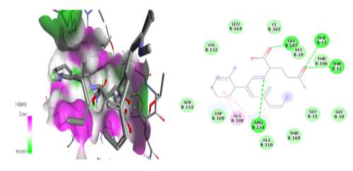
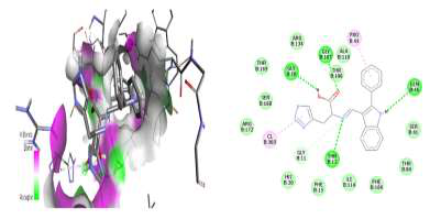
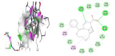
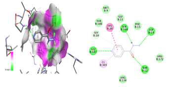
As summarized in Table 1, the molecular docking scores indicate that the synthesized Schiff base compounds, particularly 2-((2-phenyl-1H-indol-3-yl)methyleneamino)-3-methylbutanoic acid, show more negative predicted scores for the reported receptor structure than the comparator isoniazid. These scores do not, on their own, establish statistically significant differences or biological inhibitory effects. The score differences may reflect the docking algorithm’s treatment of proposed hydrogen bonds and hydrophobic contacts with receptor residues. The interaction list must be rechecked against the original PDBQT file and the matching docked complex. In particular, SER168 is incompatible with the standard residue numbering shown for the 6PUV lectin domain, and the claimed contact cannot be accepted without an independently authenticated receptor and residue-numbering map. These predicted interactions justify additional work on the ligand series, but they do not show that the derivatives are more effective inhibitors of microbial proteins than isoniazid. Their possible value as antituberculosis lead compounds remains a hypothesis requiring a verified mycobacterial target and corresponding biological evidence. However, it is important to note that molecular docking alone cannot confirm biological efficacy; additional experimental validation through in vitro and in vivo assays is essential to assess antituberculosis activity, cytotoxicity, and pharmacokinetic properties. Moreover, structure–activity relationship (SAR) studies could provide valuable insights for optimizing these compounds to enhance potency, selectivity, and drug-likeness. In conclusion, the docking results provide a preliminary computational comparison of Schiff base derivatives, subject to verification of the receptor identity and the computational setup. The combination of comparatively negative docking scores and predicted hydrogen-bonding and hydrophobic contacts supports further examination of these structures, although the calculations cannot confirm strong interactions with microbial targets. Among the tested compounds, 2-((2-phenyl-1H-indol-3-yl)methyleneamino)-3-methylbutanoic acid exhibited the highest binding affinity, making it a reasonable candidate for additional receptor-specific docking, biochemical testing, and antimicrobial evaluation before any implication for tuberculosis treatment or clinical use can be considered.
| Heterocyclic Schiff base compounds | Docking score (kcal/mol) | Predicted binding interactions |
|---|---|---|
| (E)-2-((2-phenyl-1H-indol-3-yl)methyleneamino)-4-carbamoylbutanoic acid | -7.6 | Hydrogen bonds: GLY107, PHE13, THR12, ARG134; hydrophobic contacts: ALA108 |
| (E)-2-((2-phenyl-1H-indol-3-yl)methyleneamino)-3-(1H-imidazol-4-yl)propanoic acid | -8.6 | Hydrogen bonds: GLY10, GLY107, GLN46, THR12; hydrophobic contacts: PRO44 |
| 2-((2-phenyl-1H-indol-3-yl)methyleneamino)-3-methylbutanoic acid | -8.9 | Hydrogen bonds: SER41, TRP45, GLN46; hydrophobic contacts: PRO44, ALA110 |
| Reference drug (isoniazid) | -5.9 | Reported hydrogen bonds: SER168, ASP14, THR12, GLY107; hydrophobic contacts: HIS20 |
The present study demonstrates the feasibility of combining a 2-phenylindole scaffold with amino-acid-derived Schiff-base functionality to generate structurally diverse compounds with promising predicted interactions with the reported receptor structure assigned PDB ID 6PUV. Importantly, the accession corresponds to a human lectin domain, not a Mycobacterium tuberculosis protein, and this discrepancy must be resolved before target-specific biological interpretation. Three derivatives based on L-glutamine, L-histidine, and valine were synthesized and characterized using spectroscopic and mass-spectrometric techniques, although certain formula-to-mass assignments require further confirmation. Molecular docking indicated favorable predicted binding energies of \(-7.6\), \(-8.6\), and \(-8.9\) kcal/mol for the glutamine-, histidine-, and valine-derived derivatives, respectively, compared with \(-5.9\) kcal/mol for isoniazid. Despite these encouraging computational findings, an important distinction remains between an algorithm-derived docking score, actual receptor binding, and experimentally demonstrated antitubercular activity. Docking scores cannot by themselves establish target inhibition, cellular activity, selectivity, or therapeutic efficacy. Therefore, the principal priorities for future research are in vitro antimycobacterial evaluation, cytotoxicity and selectivity studies, experimental confirmation of the proposed ligand–protein interactions, and subsequent in vivo assessment. Further research should also establish structure–activity relationships among amino-acid-derived Schiff bases and investigate how modifications of the indole scaffold and amino-acid side chains influence potency, physicochemical properties, target selectivity, and drug-likeness. Computational approaches such as molecular dynamics simulations and complementary binding-energy analyses may provide additional insight into the stability of the predicted complexes before experimental prioritization. Ultimately, integrating computational screening with biochemical, cellular, pharmacokinetic, and in vivo studies will be necessary to determine whether these compounds can progress from preliminary computational leads to validated antitubercular candidates. The valine-derived compound had the most negative of the reported docking scores and may be prioritized for further investigation; however, even the designation of a preliminary antituberculosis lead should await confirmation of a relevant mycobacterial target and experimental evidence of activity and safety. Thus, the principal significance of this work is not the demonstration of a new therapeutic agent, but the description of a structurally defined series of Schiff bases that could support subsequent receptor-verified computational studies, experimental validation, and medicinal-chemistry optimization.
Author Contributions: The authors reported equal contributions. Specific contributions should be confirmed using the journal’s required authorship taxonomy.
Conflicts of Interest: The authors reported no conflicts of interest.
Data Availability: Data are available from corresponding author upon reasonable request.
Funding Information: No funding was reported by the authors.